
Our history - Imaging platform
The IMAGING PLATFORM was established in 2021 through the initiative of the faculty and technical staff of the Anatomy and Histology Section, within the Department of Experimental and Clinical Medicine at the University of Florence.
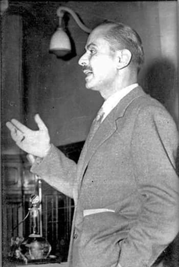
The expertise of the Imaging Platform staff in the field of morphology has deep roots. A first transmission electron microscope was installed in the early 1960s in what was then the Institute of Histology of the Faculty of Medicine and Surgery. This was strongly championed by its founder, Professor Enrico Allara, who with great foresight recognized the potential of this instrument for biomedical research.
At the turn of the last millennium, Professor Giovanni Orlandini keenly anticipated the future prospects of laser scanning confocal microscopy—which was then still in its early stages of biological application—and worked to ensure that the Department acquired one.
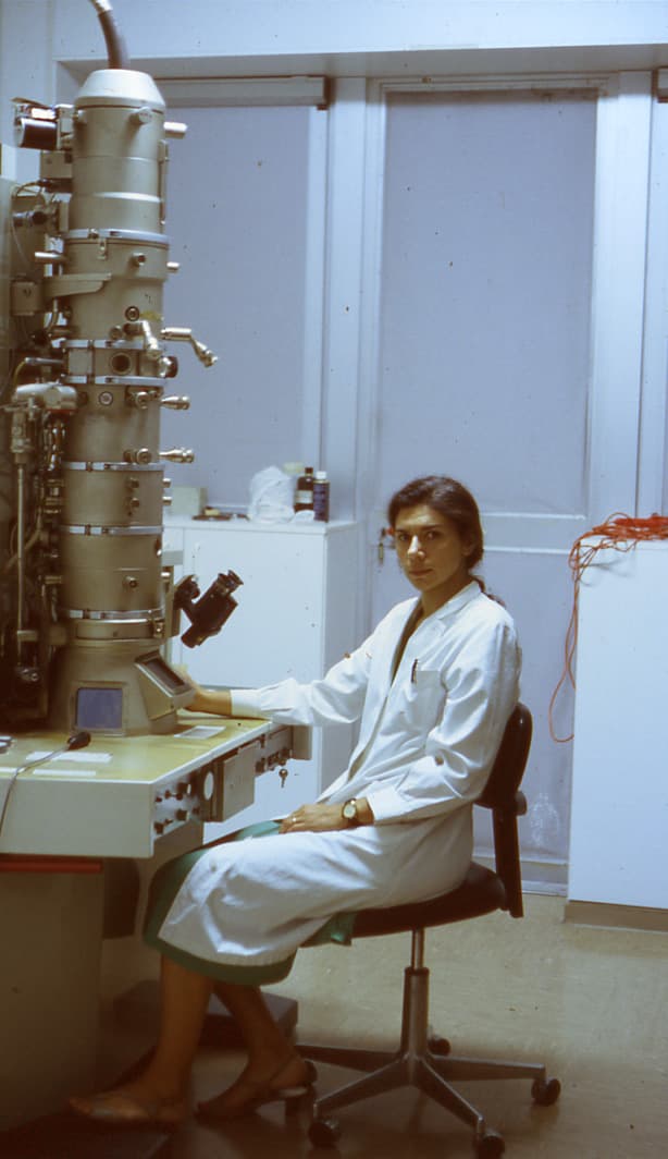
Since then, three scientific generations of researchers have succeeded one another, equipping over time a comprehensive precision microscopy laboratory that includes optical, laser confocal, and electron microscopy, as well as cell culture and molecular biology facilities. Throughout this time, they have passed down a considerable wealth of knowledge on the structure and ultrastructure of cells, tissues, and organs under normal and pathological conditions.
 |
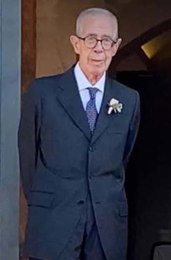 |
| Prof. Enrico Allara (1910-1985) | Prof. Giovanni Orlandini (1934-2022) |
 |
 |
 |
|
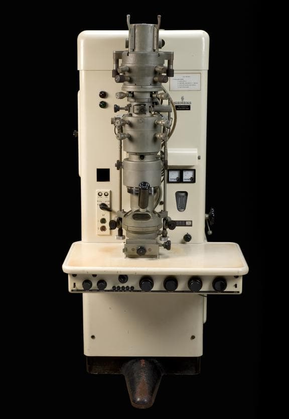
Our first Siemens Elmiskop 1A electron microscope (1963) |
Our second high-performance electron microscope, the Siemens Elmiskop 102 (1975) |
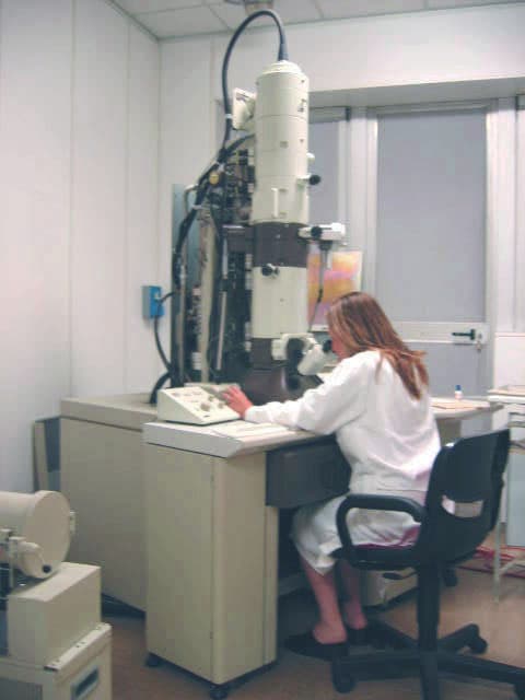
Our current digital electron microscope, the JEOL JEM-1010 (1995) |
MAIN COLLABORATIONS
University of Florence
- Department of Experimental and Clinical Medicine
- "Mario Serio" Department of Biomedical, Experimental and Clinical Sciences
- Department of Health Sciences
- Department of Neuroscience, Psychology, Drug Research and Child Health (NeuroFarBa)
- "Ugo Schiff" Department of Chemistry
Other Universities and Research Centers
- Careggi University Hospital, Florence
- Meyer Children's University Hospital, Florence
- Department of Drug Science and Technology, University of Turin
- Molecular Genetics Laboratory, Giannina Gaslini Institute, Genoa
- Carol Davila University of Medicine and Pharmacy, Bucharest, Romania
- Autonomous University of Barcelona, Spain
Institutions and Foundations
- National Research Council (CNR)
- Ente Cassa di Risparmio di Firenze (Fondazione CR Firenze)
- Menarini Foundation, Florence
- Meyer Foundation, Florence
Private Companies and Enterprises
- Janssen Research & Development
- General Project Srl., Montespertoli (Florence)
- Istituto Prosperius Srl., Florence
- Centro Odontostomatologia Laser Dott. Marco Giannelli Snc., Florence
- Symbiotec Srl., Spin-off of the University of Camerino (Macerata)
- Alcon Ltd., Switzerland
- Anatomage Italy Srl., Milan
Last update
04.06.2026