
Optical microscopy
SAMPLE PREPARATION
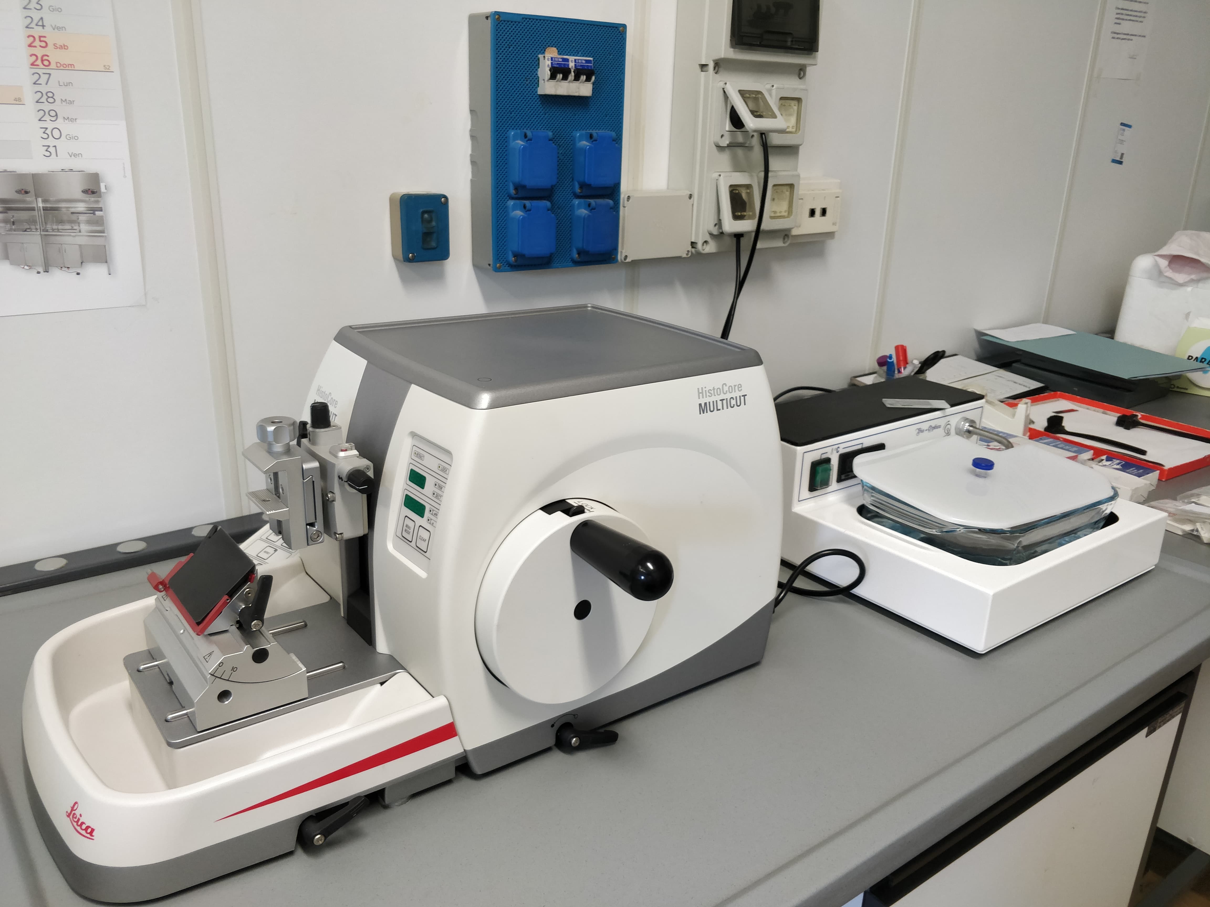
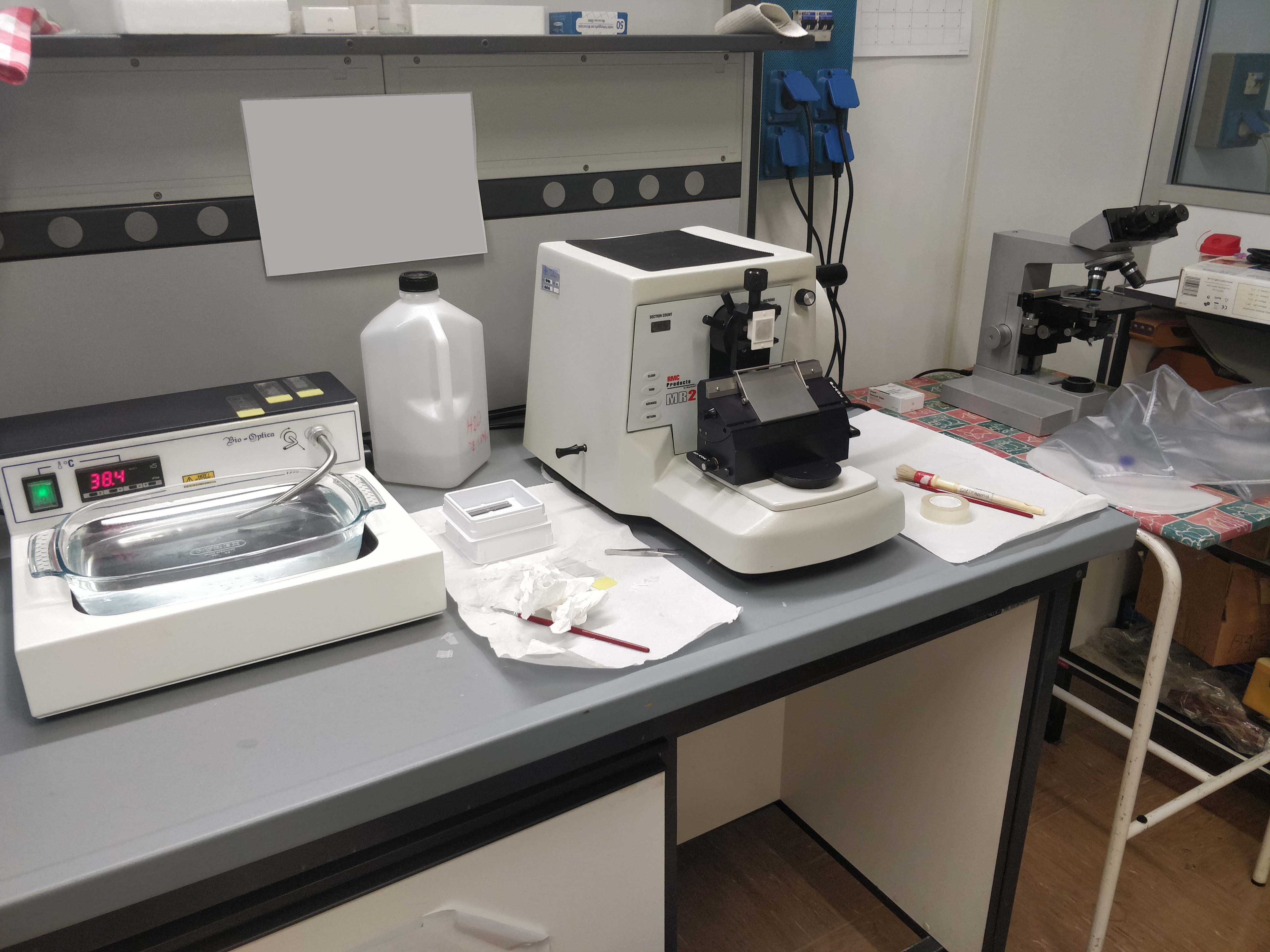
- Embedding and sectioning of paraffin-embedded tissue samples
- Embedding and sectioning of frozen tissue samples
- Histological and histochemical stainings
- Immunohistochemistry
- Immunofluorescence
.jpg) |
 |
 |
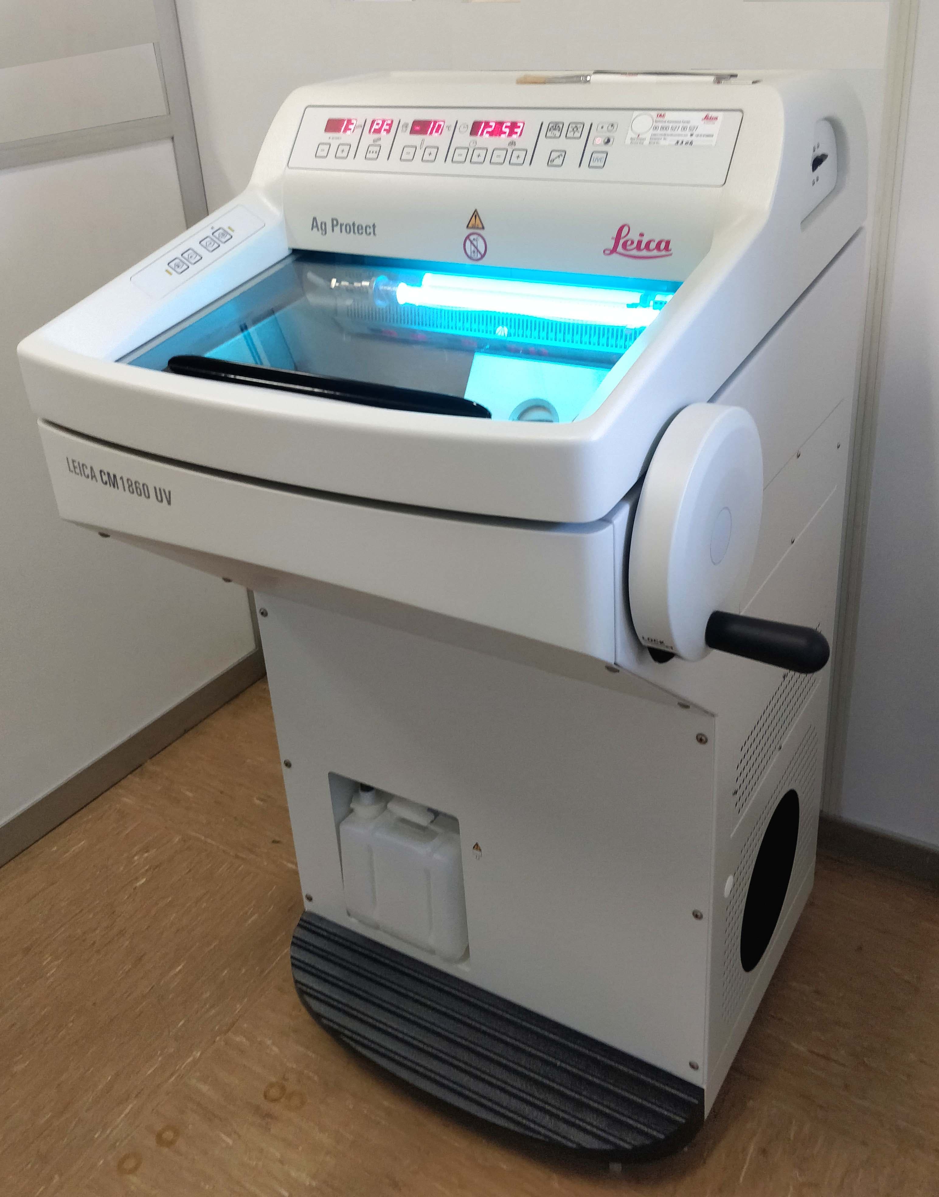 |
| Semi-automated paraffin embedding system | Microtome workstations for the preparation of thin paraffin-embedded sections | Cryostat microtome for the preparation of thin frozen sections | |
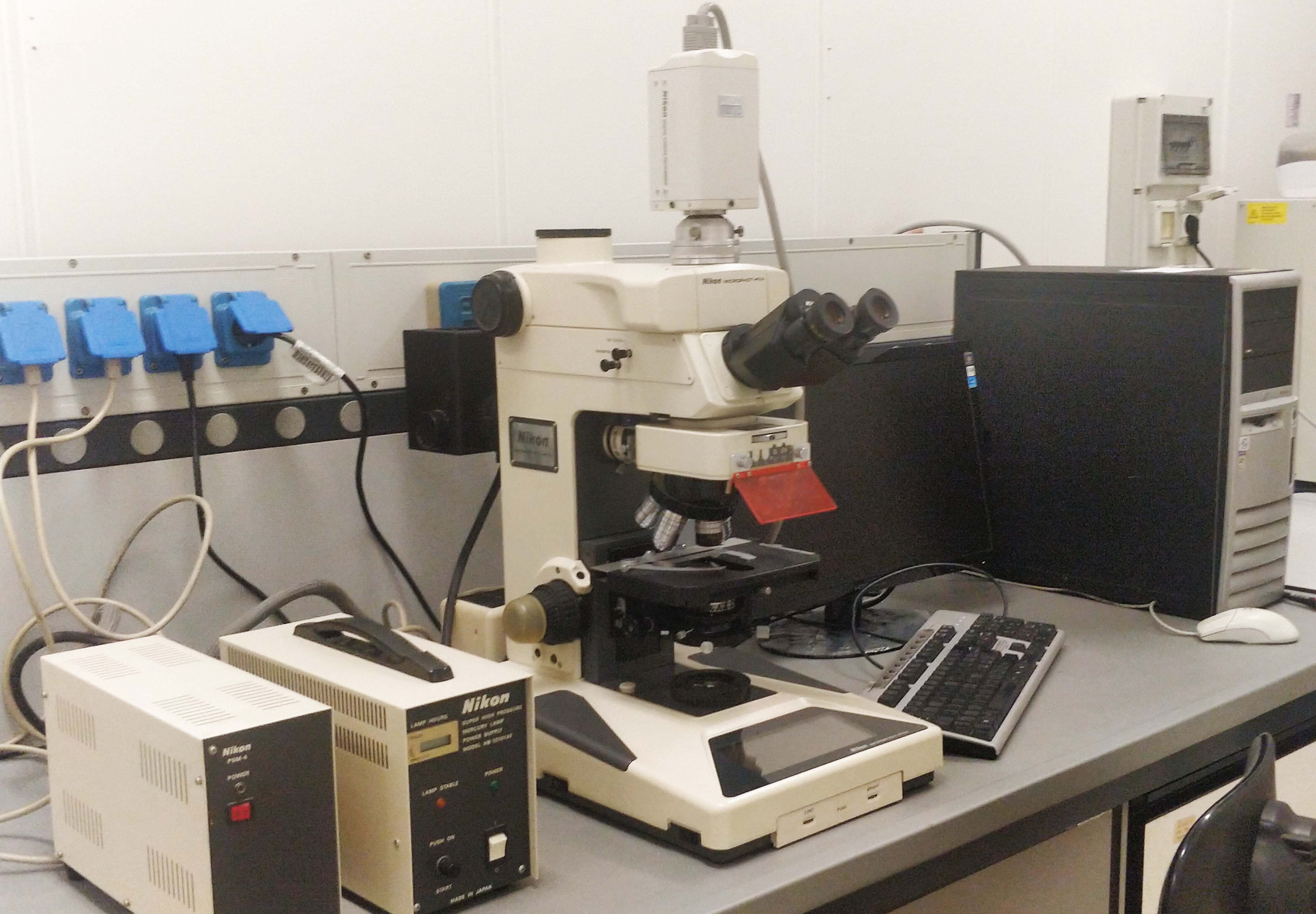
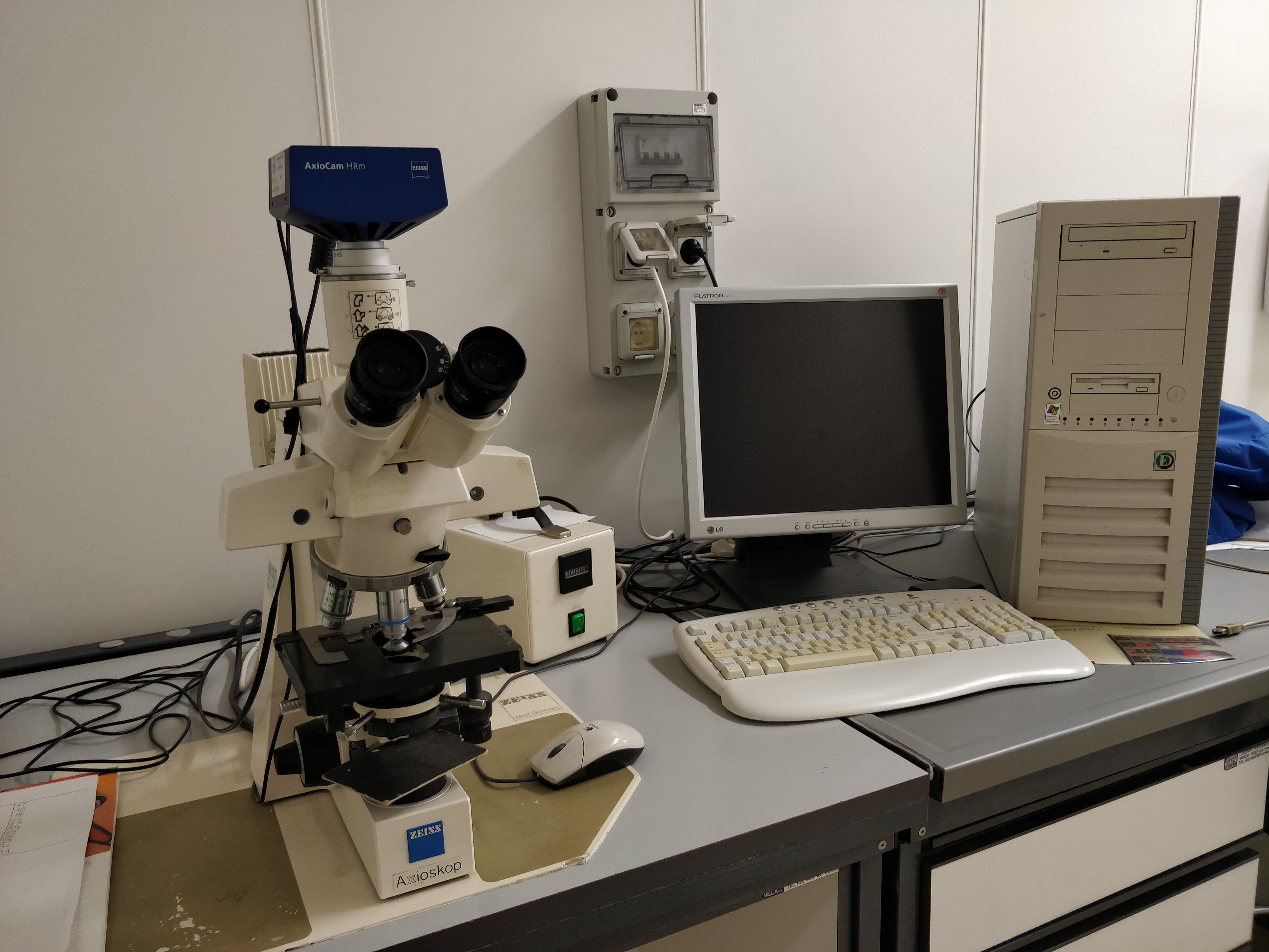
SAMPLE ANALYSIS
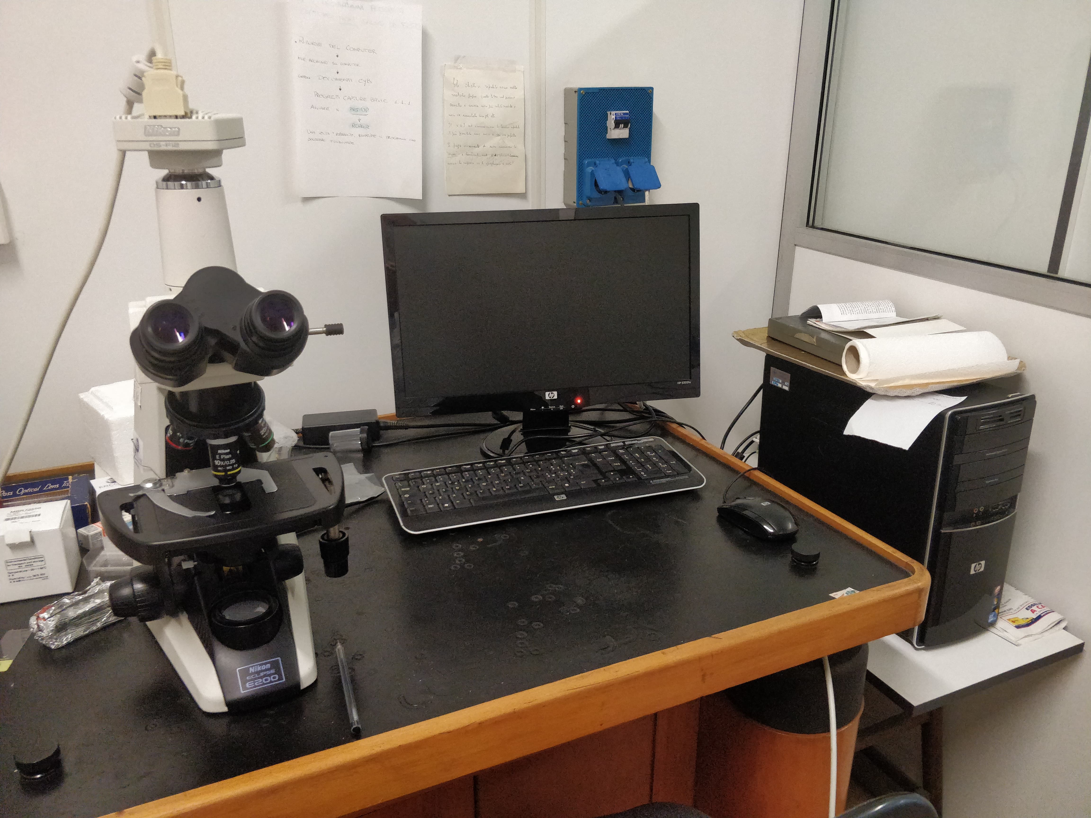
- Visible light optical microscopy
- UV light optical microscopy for fluorescence
- Digital microphotography and image analysis (ImageJ; AIVIA software): linear, area, and optical density measurements, etc.
- Histological stainings
- Histochemistry
- Immunohistochemistry
- Immunofluorescence
- Whole-slide imaging of microscopic specimens for research and diagnostics
- Morphometry
 |
 |
 |
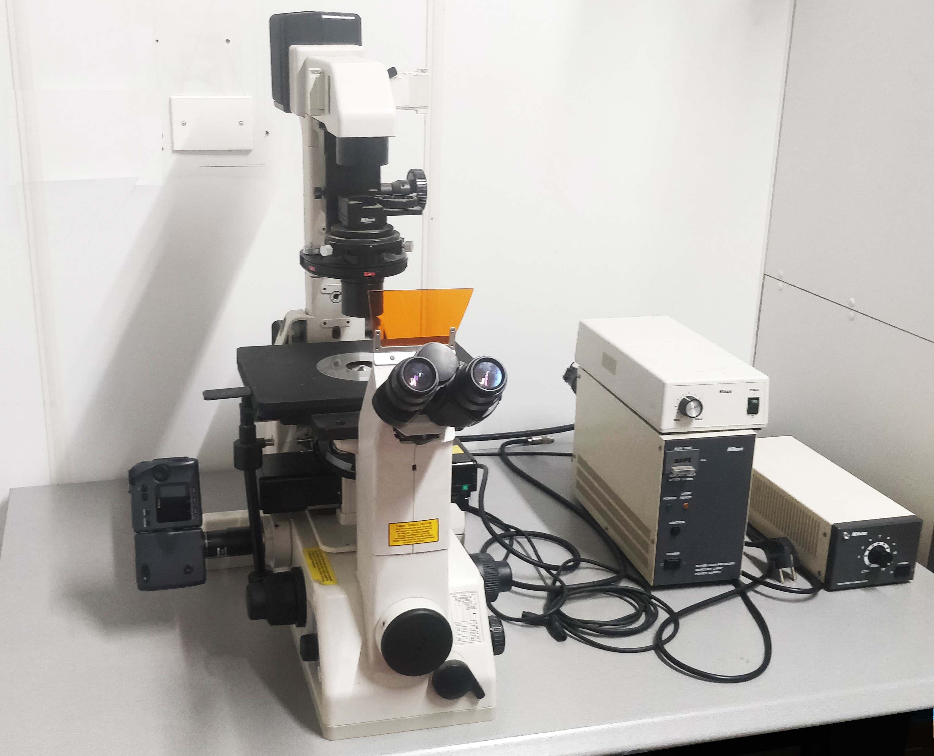 |
| Visible light optical microscopes with digital photography and image analysis systems | UV light fluorescence microscopes with digital photography and image analysis systems | Inverted microscope for visible light and fluorescence analysis of cell cultures with digital imaging system | |
 |
GRUNDIUM OCUS®40 brightfield optical microscopy slide scanner. Thanks to the excellent resolving power, chromatic aberration correction, and complete flatness of its objective lens (Olympus), it enables the high-resolution acquisition of whole-slide microscopic specimens with high color fidelity and a digital magnification of up to 800% without losing image quality. It allows for the real-time cloud archiving of high-resolution images and remote sharing via the internet with the user for research and diagnostic purposes. |
Last update
04.06.2026